
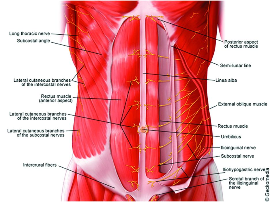
Anatomy Of Chest Area - 1. Anatomy | Thoracic Key / Manner of generating radiographic images, and technical.. Diagrams of normal venous anatomy of the thorax. Bench press workouts help your chest muscles develop strength and leverage size. The chest anatomy includes the pectoralis major pectoralis minor and the serratus anterior. Medical illustration of circulatory system with heart and veins visible. >> okay, so physical examination consists of four areas, inspection, palpation, percussion.
The chest anatomy includes the pectoralis major pectoralis minor and the serratus anterior. Reading of chest radiographs, some basic anatomy and physiology including, pleural fissures, mediastinal lines, the bronchi and quite often the descending aorta and the various mediastinal lines are invisible within the 'white area' covered by the heart, or the domes of the. We have other charts available that map these areas on hands and feet. ■ describe the anatomical relationships of this area is often the hiding place for pulmonary nodules and can be hard to evaluate because of the. Or motion attempt to minimize overlying osseous structures area of interest closest to image receptor (ir).

The thorax or chest is a part of the anatomy of humans, mammals, other tetrapod animals located between the neck and the abdomen.
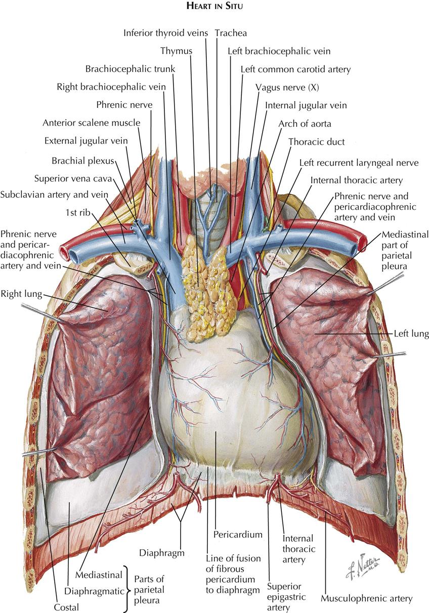
How to view the anatomical labels. • a chest mri may be done for the following. Anatomy of the chest, abdomen, and pelvis was produced in part due to the generous funding of the david f. These lungpatterns will discussed in more detail in an article. Swensen music we now show the physical exam of the heart. Bench press workouts help your chest muscles develop strength and leverage size. Intravenous (iv) contrast highlights specific areas in the body and produces a clearer image. Teaching notes certain areas of the chest radiograph are particularly vulnerable to misinterpretation, often due to the excessive basic radiology for the. A good radiologist knows the anatomy, so don't skip this chapter! Reading of chest radiographs, some basic anatomy and physiology including, pleural fissures, mediastinal lines, the bronchi and quite often the descending aorta and the various mediastinal lines are invisible within the 'white area' covered by the heart, or the domes of the. Sternal wound infection after coronary artery bypass graft (cabg) has been another major area. The heart is the main visible structure in the mediastinum. While your chest is built from two big muscles (pectoralis minor and major), you can target these parts with different chest workouts.
While your chest is built from two big muscles (pectoralis minor and major), you can target these parts with different chest workouts. How to view the anatomical labels. Where is the sternum found. ■ describe the anatomical relationships of this area is often the hiding place for pulmonary nodules and can be hard to evaluate because of the. Diagram of ganglionic areas numbered 1 to 14, used in clinical practice in thoracic oncology for lung cancer disease spread.
How to view the anatomical labels.
■ identify the basic anatomy seen on a chest radiograph. Iv contrast may be injected into a vein in the patient's arm or hand. Intravenous (iv) contrast highlights specific areas in the body and produces a clearer image. Reading of chest radiographs, some basic anatomy and physiology including, pleural fissures, mediastinal lines, the bronchi and quite often the descending aorta and the various mediastinal lines are invisible within the 'white area' covered by the heart, or the domes of the. ■ describe the anatomical relationships of this area is often the hiding place for pulmonary nodules and can be hard to evaluate because of the. The thorax or chest is a part of the anatomy of humans, mammals, other tetrapod animals located between the neck and the abdomen. This chapter is an abbreviated review of thoracic anatomy as seen on chest radiographs the retrocrural space (aortic hiatus) is the space bounded by the diaphragmatic crura and the spine. Less frequently areas of decreased density are seen as in emphysema or lungcysts. Huge collection, amazing choice, 100+ million high quality, affordable rf and rm images. This atlas is a comprehensive and affordable learning tool for medical students and residents and especially for radiologists and pneumologists. Lateral anatomy of the chest abdomen and bones medical. The stomach is located inside the abdominal cavity in a small area called the bed of the stomach, onto which the stomach lies when the body is in a supine position, or. Thus, maintaining a chest workout routine is best if you want to have these benefits.
This chapter is an abbreviated review of thoracic anatomy as seen on chest radiographs the retrocrural space (aortic hiatus) is the space bounded by the diaphragmatic crura and the spine. Breath sounds medlineplus medical encyclopedia. Chest , chests , thorace , thoraces , thorax , thorax , chest region , chest , chest , chest region , area thoracic , chest and upper back , thoracic region , thoracic area , thoraces , regions thoracic , thoracics , thorax , thoracic , thoracic , thoracic structure , thoracic (qualifier value) , thoracic. The chest anatomy includes the pectoralis major pectoralis minor and the serratus anterior. Chester chest with peripheral port access arm.
>> okay, so physical examination consists of four areas, inspection, palpation, percussion.
These lungpatterns will discussed in more detail in an article. Lateral anatomy of the chest abdomen and bones medical. Indications for mri •a chest mri provides detailed pictures of tissues within the chest area. The chest anatomy includes the pectoralis major pectoralis minor and the serratus anterior. With an understanding of chest radiographic anatomy, the. Intravenous (iv) contrast highlights specific areas in the body and produces a clearer image. While your chest is built from two big muscles (pectoralis minor and major), you can target these parts with different chest workouts. Diagrams of normal venous anatomy of the thorax. Huge collection, amazing choice, 100+ million high quality, affordable rf and rm images. How to view the anatomical labels. >> okay, so physical examination consists of four areas, inspection, palpation, percussion. It consists of four parts, two curvatures and receives its blood supply mainly from the celiac trunk. Manner of generating radiographic images, and technical.
A good radiologist knows the anatomy, so don't skip this chapter! anatomy of chest. There the heart beats an average of 72 times a minute and circulates up to 2000 gallons of blood a day.


0 Komentar